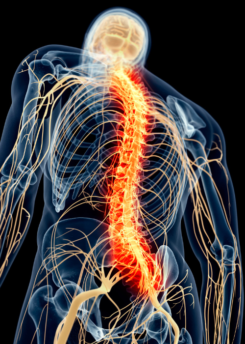
The backbone, or spine, is the most essential structure in human anatomy. The spine provides our bodies with stability, range of motion, and perhaps most importantly it keeps the major nerve root, the spinal cord, protected from pressure and outside trauma.
The spine is primarily composed of a series of 33 small bones called vertebrae. Each one separated from the others by a layer of fibrous, shock-absorbent, spongy tissue known as intervertebral discs.
This system of vertebrae and intervertebral discs form an S-shaped column that begins at the base of the neck and extends all the way down to the tailbone.
The spine is divided into five sections. The first section is called the Cervical Spine, and it is made up of the top 7 vertebrae at the neck. The next 12 vertebrae (in upper to mid back) are known as the Thoracic region of the spine. The Lumbar spine is composed of the 5 vertebrae in the lower back. Beneath these three main sections of the spine, lie the remaining two sections and 9 vertebrae in the Sacrum and Coccyx respectively.
Vertebra
Each individual vertebra is made up of two primary components; these are Vertebral Body and the Neural Arch. With the exception of the first and second cervical vertebrae, which have a slightly different structure, the remaining vertebrae all follow the same structural pattern. Each one has two vertebral laminae, two pedicles, one spinous process, two transverse and four articular processes.
The laminae form the arch itself, while the transverse processes branch out from the side. The pedicles act as anchors to which the surrounding musculature is attached, and through which movement is possible. The spinous process lies directly beneath the skin and can be felt as slight bumps all the way down the middle of the back.
- Laminae – The laminae of the vertebra can be described as broad bony plates that extend outwards from the pedicles and complete the vertebral arch.
- Spinal Canal – This spinal canal is the term used to describe the large central opening encased by the spinal arch and the vertebral body. Running through the spinal canal, we find the spinal cord and all its associated nerve roots. The primary purpose of this canal is to offer a bony armor to the delicate jelly-like substance of the spinal cord.
- Pars Interarticularis – Simply referred to as the Pars, this is where the facet joints and the back of the spine meet. The Pars Interarticularis is a bony mass and if injured, a condition known as spondylolysis may occur.
Fibrous Tissue
- Intervertebral Disc – The cartilaginous structures known as the Intervertebral Discs are located between each the vertebrae all along the spine. Each disc, or pad, forms a fibrous cushion that provides flexibility to the spine and allows for slight movements of the vertebrae.
The Intervertebral Discs also act as ligaments that hold the spine together and as shock absorbers to minimize trauma to the spinal cord.
The disc itself can be divided into two regions. The outer region is a fibrous ring called the Annulus Fibrosus. The softer, inner region has a jelly-like consistency and is dubbed the Nucleus Pulposus.
- Spinal Cord – The spinal cord is a long, thin cord of nervous tissue that runs through the length of the spinal canal. The spinal cords main function is to deliver and receive nerve impulses from the brain and to the body, and vice versa. Additionally, the spinal cord enables all function of the reflex response, as well as the central nervous system and the sympathetic nervous system.
- Facet Joint – Facet joints are the paired synovial joints that are formed by the vertebral processes and exist between each vertebra.
Facet joints provide the spine with its range of motion and a certain degree of flexibility.